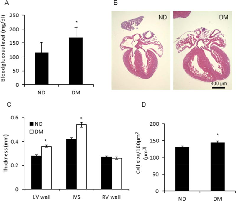
Figure 1. Cardiac hypertrophy in embryos from a T2DM dam.
A, Blood glucose level of T2DM mouse model. ND, Nondiabetes, DM, type 2 diabetes mellitus. B, Enlarged heart in embryos from ND and DM dam. C, Thickness of left ventricle (LV) wall, interventricular septum (IVS), and right ventricle (RV) wall of embryo from ND and DM dam. D, Cardiomyocyte size in heart of embryo from ND and DM dam. 76 and 40 hearts of embryos from 16 ND dams and 40 DM dams, respectively, were removed at E17.5. * indicates significant difference (P < 0.05) when compared to ND group.

