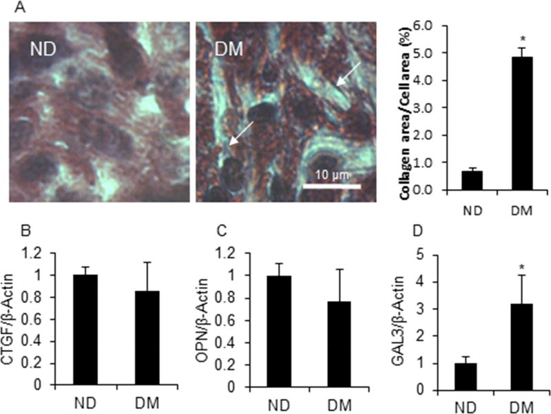
Figure 5. T2DM induced pro-fibrosis in the embryo heart.
A. Masson staining of the embryo heart section. Arrow represents collagen staining (blue line). mRNA expression of pro-fibrosis markers, CTGF (B), OPN (C), and GAL3 (D). Each group contained three samples. * indicates significant difference (P < 0.05) when compared with the ND group.

