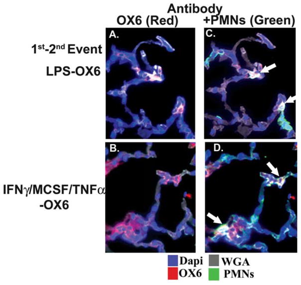
Fig. 4.
OX6 colocalizes with the polymorphonuclear neutrophil (PMN) membranes. Rats received an intraperitoneal injection of either: 1) interferon-γ (IFNγ) and macrophage-colony–stimulating factor (MCSF) 24 hours before the infusion and intravenous TNFα 2 hours before the infusion, or 2) 2 mg/kg lipopolysaccharide (LPS) 2 hours before the infusion of OX6. After 6 hours, the rats were killed; and the left lung was inflated with optimal cutting temperature compound, embedded in paraffin, frozen, and sectioned. The sections were incubated with wheat germ agglutinin (WGA) (gray), which localizes to the cellular membrane (gray); 4′,6-diami-dino-2-phenylindole (DAPI) (blue), a nuclear stain; an antibody to OX6 (red); and an antibody specific for rat PMNs (green). All sections were visualized at ×40 magnification in duplicate. (A) OX6 (red) colocalized to cells, which were PMNs (green), as demonstrated in C, and this “triple colocalization” was present on the PMN membrane (white), as demarcated by white arrows. (B) Antibodies to OX6 (red) were most widespread, which were localized to both the PMNs (green) with triple colocalization to the PMN membrane (white, white arrows) in D. (C,D) There is some OX6 on the lung tissues (red) that is not associated with PMNs, although most of it is associated with infiltrating PMNs. Such immunoreactivity may be expected in rats treated with IFNγ/MCSF/TNFα, and especially IFNγ, which induces OX6 surface expression on both lung endothelium and epithelium.31–33

