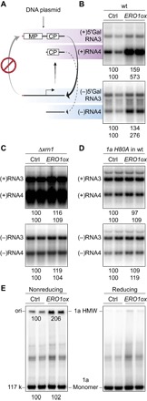
Fig. 2. Ero1p stimulates BMV (+)RNA accumulation by enhancing 1a’s disulfide-linked complex formation and capping function.

(A) Schematic representation of the limited BMV-directed RNA synthesis pathway for the RNA3 derivative 5′ Gal RNA3, which has the WT 5′ untranslated region replaced with that of yeast GAL1 mRNA (red line). MP (movement protein) and CP (capsid protein) designate RNA3 open reading frames. (B) Analysis of positive- and negative-strand 5′ Gal RNA3 and RNA4 from WT yeast cells expressing 1a, 2apol, and either vector (ctrl) or exogenous Ero1p (ERO1ox). (C) Analysis of RNA3 and RNA4 from Δxrn1 yeast cells expressing 1a, 2apol, and either vector or exogenous Ero1p. (D) Analysis of RNA3 and RNA4 from WT yeast cells expressing a capping-defective mutant 1a H80A, 2apol, and either vector or exogenous Ero1p. (E) Analysis of disulfide-linked 1a complexes from WT yeast cells used in Fig. 1B. 1a monomer and dithiothreitol (DTT)–sensitive, high–molecular weight (HMW) complex were analyzed by SDS–polyacrylamide gel electrophoresis (SDS-PAGE) using neutral 3 to 8% SDS-PAGE gels under both reducing and nonreducing conditions.
