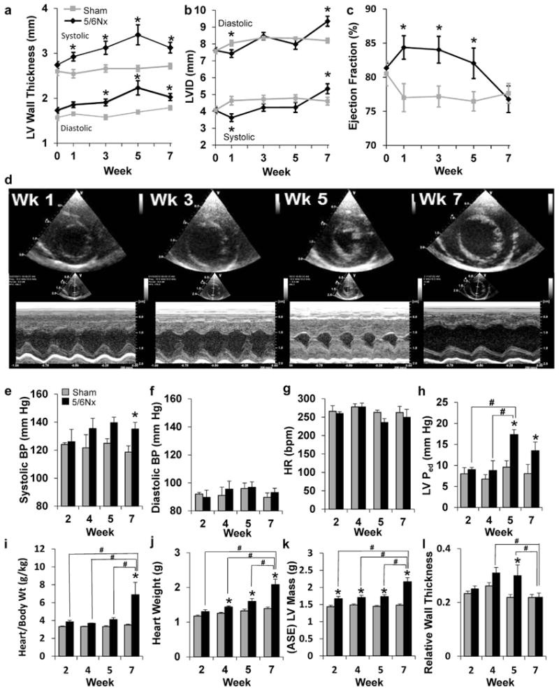
Figure 2. Indices of left ventricular (LV) remodeling and function.

(a) Measurements of wall thickness, (b) LV inner diameter (LVID), and (c) percentage of ejection fraction were made from M-mode echocardiography images before surgery and at weeks 1, 3, 5, and 7 post-surgery. N = 12 per group; *P < 0.05 versus sham controls; 2-way repeated-measures analysis of variance. (d) Representative echocardiograms of end-diastolic dimension show dramatic changes inboth wall thickness and chamber size inresponse to 5/6 nephrectomy (5/6Nx) (upper, short-axis view; lower, M-mode view). Hemodynamic influences on the left ventricle were measured in animals before tissue collection for gene expression analysis at weeks 2, 4, 5, and 7 post-5/6Nx. (e) Systolic blood pressure (BP) was modestly, but significantly, increased at week 7, whereas (f) diastolic pressure and (g) heart rate (HR) remained unchanged (N = 5–6 per group). (h) Diastolic pressure in the left ventricle (LVPed) is significantly elevated at 5 and 7 weeks post-5/6Nx, suggesting diastolic dysfunction. Body weight (Wt), (i) normalized heart weight, and (j) isolated heart weight were significantly elevated as early as 2 weeks post-5/6Nx. Mass calculated (ASE) from echocardiography measurements of the LV (k) also indicated progressive hypertrophy. Relative wall thickness (diastolic LV wall thickness/diastolic chamber diameter) was significantly elevated at 4 and 5 weeks post-5/6Nx, indicating hypertrophic LV remodeling (l). N = 4–6 per group; *P < 0.05 versus sham control at time point. #P < 0.05 versus indicated 5/6Nx group, 2-way analysis of variance. ASE, American Society of Echocardiography; bpm, beats per minute; Wk, week. To optimize viewing of this image, please see the online version of this article at www.kidney-international.org.
