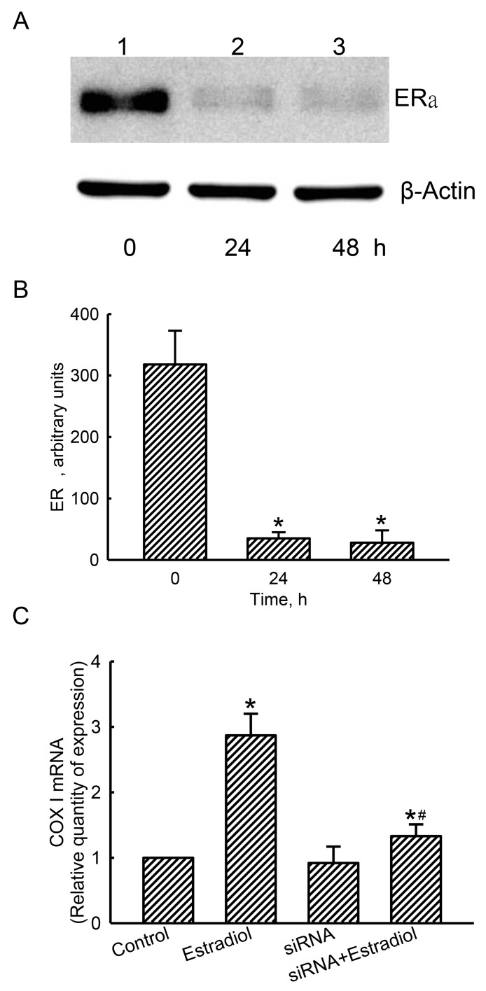
Figure 8. Effects of estrogen receptor alpha (ERα) knockdown on estradiol-induced mitochondrial cytochrome c oxidase (COX) I mRNA expression.
Human osteoblast-like U2OS cells were treated with ERα siRNA for 24 and 48 h. Scrambled siRNA was administered to control cells as the negative standard. Levels of ERα were immunodetected (A, top panel). Amounts of β-actin were analyzed as the internal standard (bottom panel). These protein bands were quantified and statistically analyzed (B). After knocking-down ERα translation for 24 h, human osteoblasts were treated with estradiol for another 6 h. A quantitative PCR analysis was conducted to determine COX I mRNA expression (C). Each value represents the mean ± SEM, n = 3. The symbols * and # indicate that a value significantly (p < 0.05) differed from the control and estradiol-treated groups, respectively.

