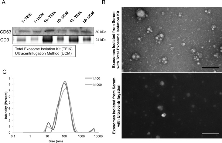
Figure 1. Exosome isolation method.
Representative CD63 immunoblot for serum exosome lysates (SEL) from identical volumes of serum from untreated SKH-1 mice, where the Total Exosome Isolation Kit (Invitrogen) (TEIK) was compared to the Ultracentrifugation Method (UM). Number indicates mouse identification number (A). Representative electron micrographs of samples isolated by either the total exosome isolation kit or ultracentrifugation showed intact exosomes of the correct size in both samples. Although, a higher level of background protein staining was observed in the samples isolated with the ultracentrifugation method, as seen here by the dark background, the scale bar is 500nm (B). Serum exosome samples were ran on the Zetasizer to determine the particle sizes present, each curve representing various dilutions (C).

