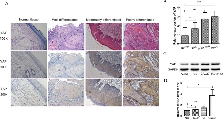
Figure 1. YAP was highly expressed in oral squamous cell carcinoma.
(A) Representative images of YAP immunostaining in normal tissues, well differentiated OSCC tissues, moderately differentiated OSCC tissues and poorly differentiated OSCC tissues. Low power (100×) scale bars 100 μm, high power (200×) scale bars: 50 μm. (B) Statistical relative quantification of YAP expression. (C) Western blot analysis of expression of YAP in four OSCC cell lines. (D) RT-PCR analysis of YAP expression in four OSCC cell lines. *p < 0.05, **P < 0.01, ***p < 0.001.

