
Figure 2. Fluorescence imaging of a primary uterine serous adenocarcinoma and of the metastatic lymph nodes.
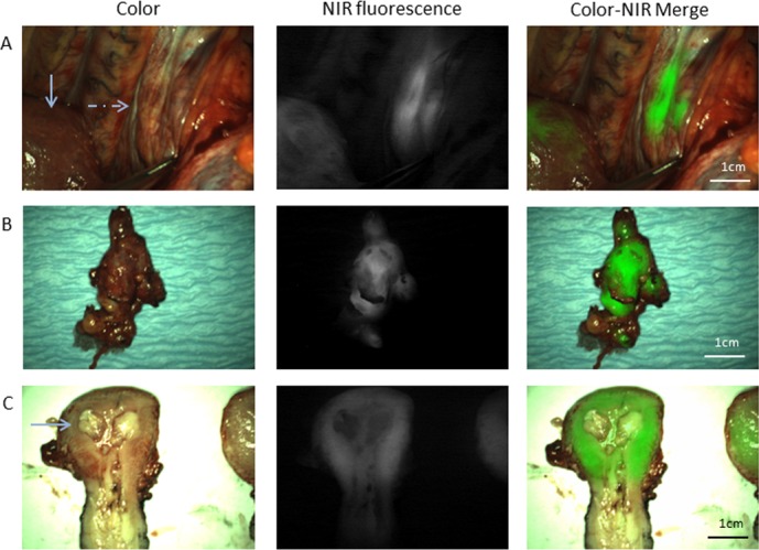
(A) Intraoperative identification of para-aortic, metastatic lymph nodes (dashed arrow), located beneath a layer of overlying tissue (patient #1). The normal arrow indicates the fluorescence signal arising from the uterus. (B) Ex vivo fluorescence imaging of the resected para-aortal lymph nodes (patient #1), that show a clear fluorescence signal. (C) Ex vivo fluorescence imaging of the bisected uterus (patient #1). The fluorescence signal, detected during surgery, appears to be mainly arising from normal adjacent background uterine tissue instead of the primary tumor.