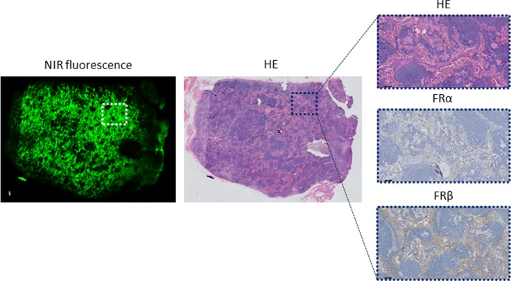
Figure 4. Histopathological evaluation of a false-positive lymph node.

Shown are a NIR fluorescence image, haematoxylin & eosin (HE) staining, and FRα and FRβ staining of a (fluorescent) LN that did not contain tumor cells. Fluorescence is mainly seen in the sinuses and not in the follicles. The magnified images (HE, FRα and FRβ) show the lack of FRα staining, while the sinuses show a positive FRβ staining. Magnifications: 1× and 10×.
