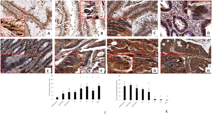
Figure 1. FZD-10 expression in hyperplastic, dysplastic and carcinomatous colon tissues from patients with sporadic cancer.
Representative images of the immuno-histochemical expression of FZD-10 in hyperplastic mucosa (A), adenomas with low-grade dysplasia (B) and high-grade dysplasia (C), in carcinomatous tissue, T1 (D), T2 (E), T3 (F), T4 (G) and metastases (H) (×40 magnification). Insets show magnified nuclei for selected areas (squares). The diagram in (J, K) shows the percentage of FZD-10 positive pixels per area in J reported as the mean ± SD obtained for the colon tissue of 19 patients and in K the % of positive nuclei in 10 different area of sample: (J) protein expression in cytoplasm and membranes, (K) Nucleic protein expression. Significance was determined by ANOVA test [p < 0.001(*) and p = 0.05] by the Holm–Sidak method.

