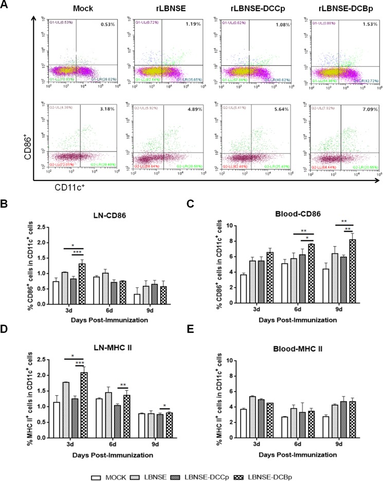
Figure 3. DC activation in mice immunized with different rRABVs.
BABLB/c mice were immunized with 1 × 10⁶ FFU of rRABVs or DMEM. The lymph nodes (LN) and blood samples were collected at 3, 6 and 9 dpi. Single cell suspensions prepared from the lymph nodes and blood were analyzed for the presence of DCs (CD11c+ and CD86+, or CD11c+ and MHC II+). (A) Representative gating strategy for DCs in blood or inguinal lymph samples. (B) and (C) Percentages of CD11c+ and CD86+ activated DCs in LN and blood samples of immunized mice respectively. (D) and (E) Percentages of CD11c+ and MHCII+ activated DCs in LN and blood samples of immunized mice respectively. Data are the means from three independent experiments (*P < 0.05; **P < 0.01; ***P < 0.001).

