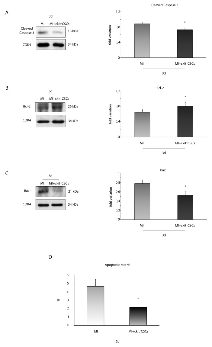
Figure 10. ckit+CSCs modulate the expression of apoptotic markers in treated infarcted hearts.
Western blot analysis showing the expression of (A) cleaved-caspase 3, (B) Bcl-2 and (C) Bax, in the border zone of 3 day-infarcted control hearts (MI, n=3) or ckit+CSC transplanted hearts (MI+ckit+CSCs, n=3). Left panel: A representative Western blotting of three independent experiments is shown. Right panel: Densitometric analysis of Western blot. Data are shown as means ± SEM. *P < 0.05 vs MI. (D) Cardiomyocyte apoptosis measured by TdT assay. Bar graph showing apoptotic rate in the peri-infarct area of the myocardium of control (MI) and ckit+CSC transplanted infarcted (MI+ckit+CSCs) hearts. Data represent means±SD (MI, n=5; MI+ckit+CSCs, n=5; *p<0.0052 vs MI).

