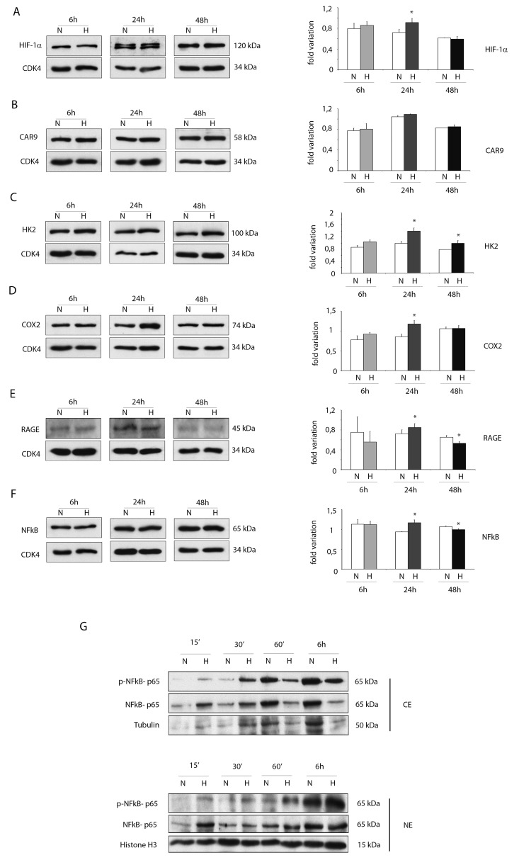
Figure 3. Hypoxia enhances protein expressions of pro-inflammatory proteins in ckit+CSCs.
Western blot analysis showing the expression of (A) HIF-1 α, (B) CAR9 and (C) HK2, (D) COX2 and (E) RAGE and (F) NFkB in ckit+CSCs under hypoxic conditions (H) at 6, 24 and 48h compared to normoxic conditions (N). The same filter was probed with anti-CDK4 pAb to show the equal loading. Left panel: A representative Western blotting of three independent experiments is shown. Right panel: Densitometric analysis of Western blot. Data are shown as means ± SEM. *P < 0.05 vs normoxic conditions (N). (G) Western blot analysis showing the expression of phospho-specific anti-NFkB p65, anti-NFkB p65, tubulin and histone H3. The analysis was performed using cytoplasmic (CE) and nuclear (NE) extracts prepared from ckit+CSCs under hypoxic conditions (H) at 15’, 30’, 60’ and 6h compared to normoxic conditions (N). A representative Western blotting of three independent experiments is shown.

