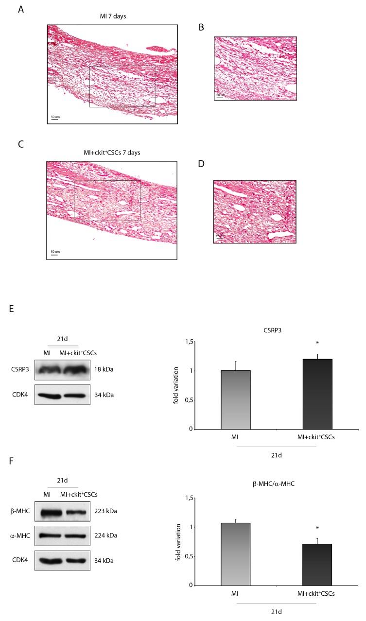
Figure 8. ckit+CSCs attenuate hypertrophy following acute MI.
Representative H&E stained myocardial sections (A-D) of 7 day infarcted region from control (MI) (A) and ckit+CSC transplanted hearts (C). (B) and (D) represent high magnification of the insets shown in (A) and (C), respectively. The relative levels of (E) CSRP3 and (F) β-MHC/ α-MHC were investigated in the border zone of 21 day-infarcted control hearts (MI, n=3) or ckit+CSC transplanted hearts (MI+ckit+CSCs, n=3) by Western blot (left panel) and relative densitometry of three independent experiments (right panel). Data are shown as means ± SEM. *P < 0.05 vs MI.

