FIGURE 6.
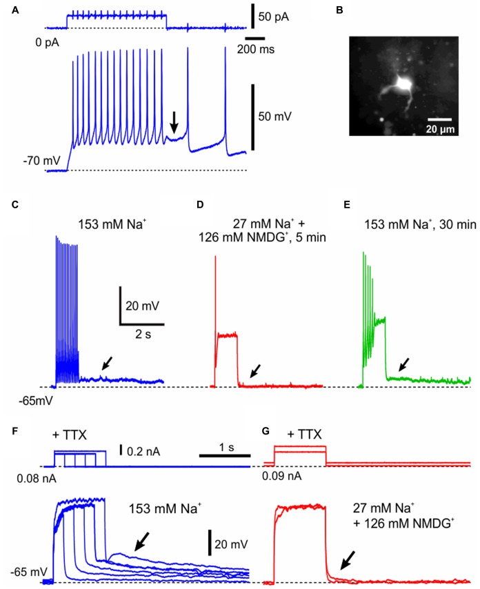
The generation of pulse-ADP is dependent on extracellular Na+. (A) An application of a depolarizing current pulse in a neuron at the resting membrane potential of –70 mV caused a spike train, which was followed by a pulse-ADP (arrow) that triggered further spikes. (B) A lucifer yellow image of the recorded neuron classified as γMN, which displayed sparse arborizations of primary dendrites. (C) A spike train that was followed by a pulse-ADP lasting for more than 5 s (arrow) was induced in aCSF containing 153 mM Na+. (D) Abolishment of the pulse-ADP (C, arrow) by substitution of 126 mM Na+ with the equimolar NMDG+. (E) Recovery of the pulse ADP (arrow) after washout of NMDG+ with the original aCSF. Compare (E) with (C,D). (F) In the presence of 1 μM TTX, the amplitude of pulse-ADPs increased (arrow) as the duration or the amplitude of the depolarizing current pulse increased. (G) In the presence of TTX, the pulse-ADP was abolished (arrow) by substitution of 126 mM Na+ with the equimolar NMDG+.

