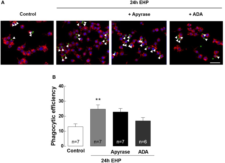
Figure 7.
ATP and adenosine do not contribute to the increase of phagocytic efficiency in microglia induced by elevated hydrostatic pressure. Phagocytosis was assessed using fluorescent microbeads. (A) Representative images of BV-2 cells stained with phalloidin (red) with incorporated beads (green). Nuclei were counterstained with DAPI (blue). Arrowheads show some beads engulfed by microglia. (B) Phagocytic efficiency. Scale bar: 50 μm. **p < 0.01, different from control; Kruskal-Wallis test, followed by Dunn's multiple comparison test.

