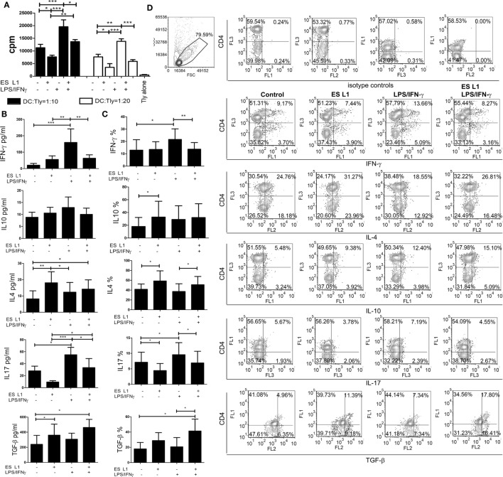
Figure 3.
Allo-stimulatory and T helper polarizing capacity of ES L1-pulsed dendritic cells (DCs). (A–D) DCs treated with ES L1 antigens (50 µg/ml) and/or LPS/interferon (IFN)-γ were washed thoroughly and then cocultured with magnetic-activated cell sorting-purified allogenic T cells (Tly) (1 × 10⁵/well) for 6 days in two DC:T cell ratios (1:10 and 1:20). (A) The proliferation in cocultures was measured by 3H-thymidin incorporation assay, and Tly cultivated alone were used as a blank control. (B) The concentration of indicated cytokines were determined in the supernatants of PMA/ionophore-treated DC:T cell cocultures at 1:20 cell-to-cell ratio, respectively, by specific ELISA tests. (C) The percentage of cytokines expression measured intracellularly by flow cytometry, within the T cells cocultivated with DCs as in (B) and treated with PMA/Ionophore/monensin for the last 4 h, are shown as mean% ± SD of four experiments with different DCs donors. *p < 0.05, **p < 0.01, ***p < 0.001 compared to control, or as indicated by line (one-way ANOVA with Tukey’s posttest). (D) The analysis of the intracellular cytokines in T cells was carried out after the surface staining of CD4, as indicated on representative dot plots collected from two experiments.

