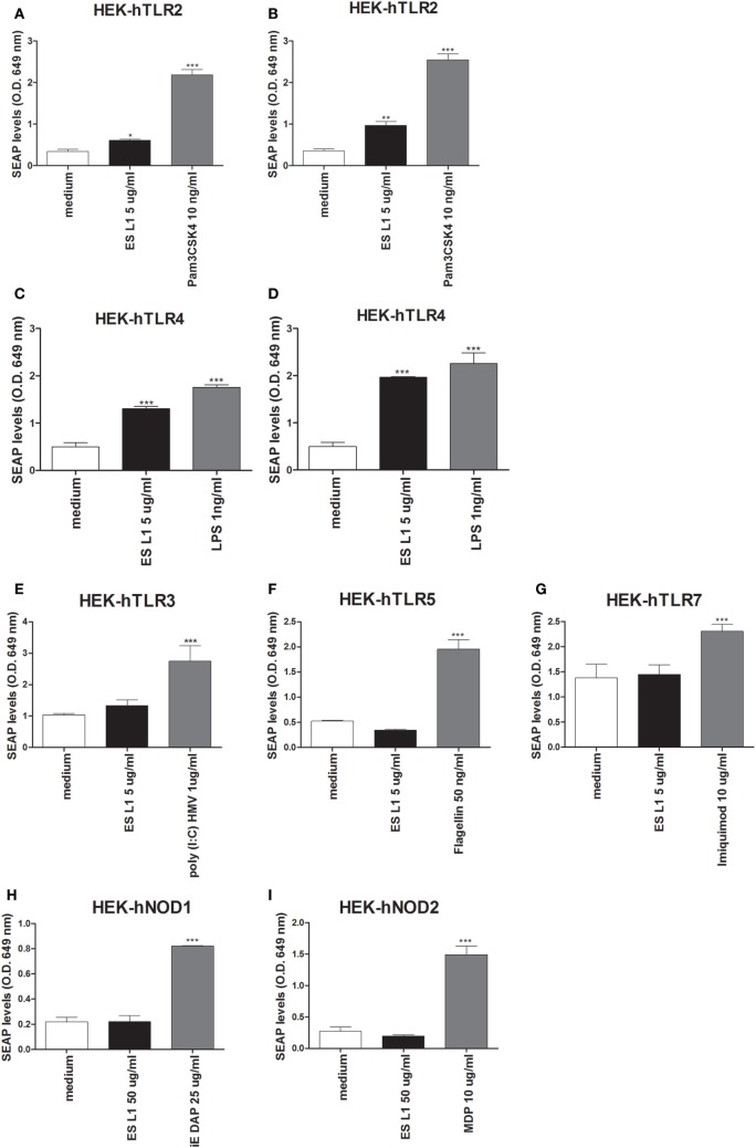
Figure 6.
Interaction of ES L1 antigen with pattern recognition receptors (PRRs) on HEK-Blue™ cell lines. HEK-Blue™ cell lines transfected with a single specific human PRR (TLR2, 3, 4, 5, 7, NOD1, and 2) were treated with ES L1 or PRR agonists for 24 h, followed by the analyses of secreted alkaline phosphatase (SEAP) levels (OD 649 nm) released in culture medium at two time points (4 and 24 h after the substrate addition). Pam3CSK4 (10 ng/ml), ES L1 (5 µg/ml)—incubation period 4 (A) and 24 h (B); LPS (1 ng/ml) Escherichia coli K12, ES L1 (5 µg/ml)—incubation period 4 (C) and 24 h (D); Poly (I:C) HMV (1 µg/ml), ES L1 (5 µg/ml)—incubation period 24 h (E); FLA-ST (50 ng/ml), ES L1 (5 µg/ml)—incubation period 24 h (F); imiquimod (10 µg/ml), ES L1 (5 µg/ml)—incubation period 24 h (G); iE-DAP (25 µg/ml) ES L1 (50 µg/ml)—incubation period 24 h (H); MDP (10 µg/ml), ES L1 (50 µg/ml)—incubation period 24 h (I). Results are shown as mean ± SD from three different experiments *p < 0.05, **p < 0.01, ***p < 0.001 compared with control (medium) (one-way ANOVA with Tukey’s posttest).

