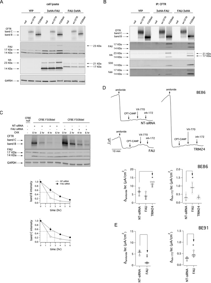
Figure 6.
Analysis of CFTR interaction with FAU. A, biochemical analysis of CFTR and FAU expression pattern in whole lysates from CFBE41o− cells after transfection with a vector coding for the YFP or vectors coding for two different FAU constructs having a triple HA tag at the N or C terminus (3xHA-FAU and FAU-3xHA, respectively). B, whole lysates used in A were immunoprecipitated using an anti-CFTR antibody. Immunoblot detection of FAU protein was performed using three different antibodies targeting different domains of the protein and an anti-HA antibody. C, upper panel, immunoblot detection of mutant CFTR in whole lysates derived from CFBE41o− cells transfected with indicated siRNA (final concentration, 30 nm) and at different time points following cycloheximide (CHX)-induced block of protein synthesis. Lower panels, quantification of mutant CFTR (band B and band C) half-life. D, representative traces and scatter dot plots summarizing data from Ussing chamber recordings of human primary bronchial epithelia from a homozygous F508del patient (BE86) following reverse transfection with 50 nm of indicated siRNA molecules. E, scatter dot plots summarizing data from Ussing chamber recordings of human primary bronchial epithelia from a homozygous F508del patient (BE91) following reverse transfection with 50 nm of indicated siRNA molecules.

