
Figure 6.
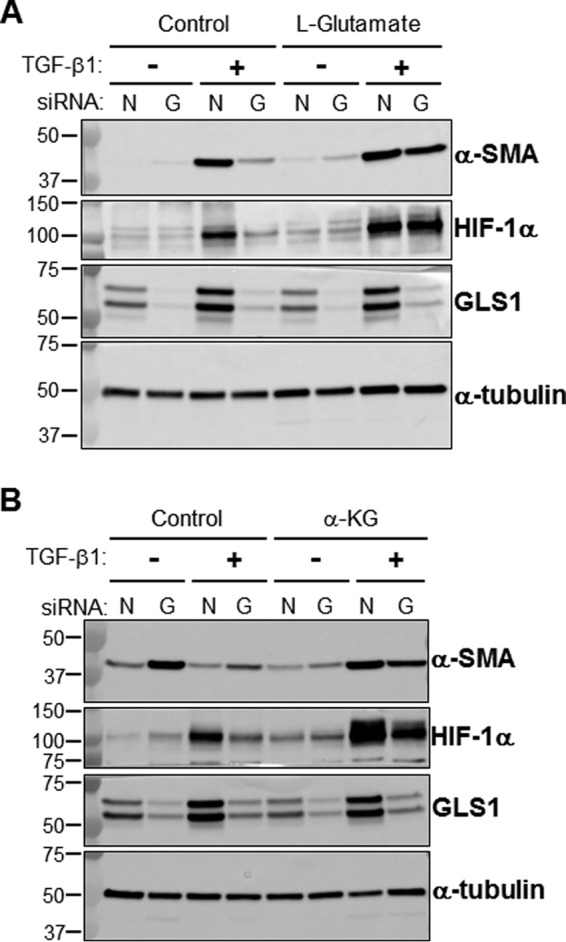
Exogenous glutamate or α-KG restores the expression of profibrotic markers in GLS1-deficient myofibroblasts. A, total cell lysates were isolated from lung fibroblasts transfected with NT (lanes N) or GLS1 (lanes G) siRNA (100 nm), treated with or without l-glutamic acid (2 mm), and treated with or without TGF-β1 (2.5 ng/ml, 48 h). l-Glutamic acid was added in the reduced serum DMEM (0.5% fetal calf serum supplemented with 2 mm l-glutamine) 1 h prior to the addition of TGF-β1. Then protein levels of α-SMA, HIF-1α, GLS1, and α-tubulin were determined by WB; molecular mass markers are indicated on the left side of the panel (representative WB of n = 3). B, total cell lysates were isolated from lung fibroblasts transfected with NT or GLS1 siRNA (100 nm), treated with or without diethyl-2-oxopentanedioate (esterified form of α-KG, 2 mm), and treated with or without TGF-β1 (2.5 ng/ml, 48 h). Diethyl-2-oxopentanedioate was added in the reduced serum DMEM (0.5% fetal calf serum supplemented with 2 mm l-glutamine) 24 h prior to the addition of TGF-β1. Then protein levels of α-SMA, HIF-1α, GLS1, and α-tubulin were determined by WB; molecular mass markers are indicated on the left side of the panel (representative WB of n = 3).