Figure 2.
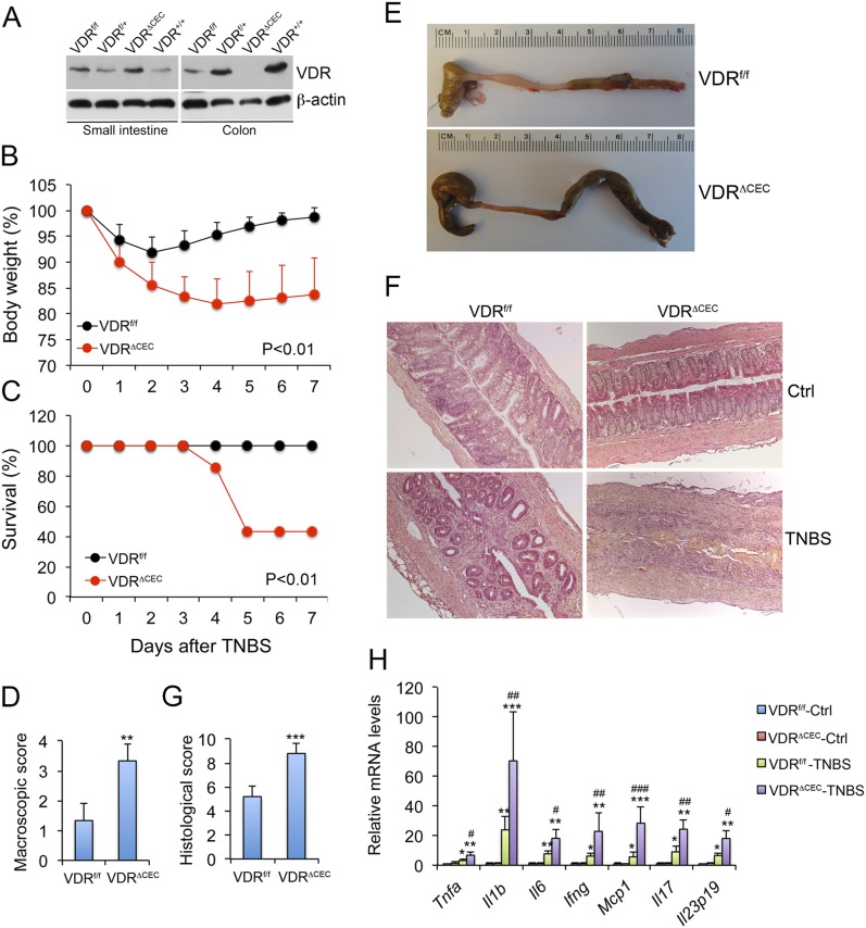
Colonic epithelial VDR deletion aggravates colonic inflammation in TNBS-induced colitis model. (A) Western blots showing VDR expression in the small intestine but not in the colon of VDRΔCEC mice. (B) Body weight changes following TNBS treatment; n = 6 to 8 mice; P < 0.01 by log rank test. (C) Survival curve; n = 6 to 8 mice; P < 0.01 by log rank test. (D) Scores of colonic macroscopic damage. (E) Gross images of the large intestine on day 7. (F) Hematoxylin and eosin staining of distal colon sections cut longitudinally on day 4. (G) Histological scores of the colons; **P < 0.01; ***P < 0.001 vs. VDRf/f; n = 4 to 5 mice. (H) Real-time RT-PCR quantitation of mucosal proinflammatory cytokines and chemokines in VDRf/f and VDRΔCEC mice with or without TNBS treatment on day 4. *P < 0.05; **P < 0.001; ***P < 0.001 vs. corresponding control; #P < 0.05; ##P < 0.01; ###P < 0.001 vs. VDRf/f-TNBS; n = 3 to 5 mice. Ctrl, control; Ifng, interferon γ; Il1b, interleukin 1B; Il6, interleukin 6; Il17, interleukin 17; Mcp1, monocyte chemotactic protein-1; Tnfa, tumor necrosis factor α.

