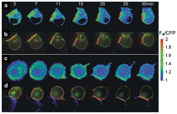
Fig. 1.
CD4 co-receptor recruitment to the immunological synapse and FRET between TCR and CD4. (a), (b) show a time course of interaction between T cell and an APC presenting antigenic peptide. (c), (d) show the same with an APC that does not present the antigenic peptide. (a), (c) show the FRET response between CD3ζ–CFP and CD4–YFP, using a heat scale (Zal et al. 2002). (b), (d) show the fluorescence of the CD3ζ–CFP (green) and CD4–YFP (red). Only the antigenic stimulation causes close interaction between TCR and CD4, as reported by FRET between CD3ζ–CFP and CD4–YFP (a versus c), though both APCs recruited CD4 to the immunological synapse (b and d). Recruitment was much slower in the absence (d) versus the presence (d) of antigen. Reproduced with permission from Zal et al. (2002)

