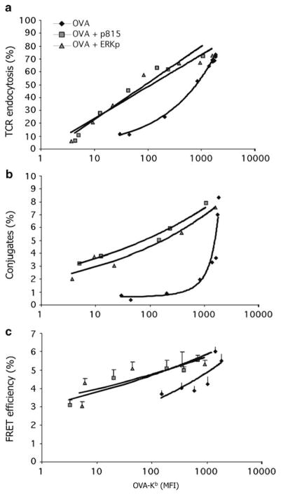
Fig. 2.
Increased T-cell activation by endogenous nonstimulatory peptides at limiting antigen quantities. (a) shows the amount of TCR endocytosis at differing quantities of antigen OVA–Kb expressed on the cell surface of RMA-S cells, either alone or with added nonstimulatory peptides derived from VSV, Erk, or the P815 tumor antigen. Erk and P815 are natural endogenous Kb–binding peptides (Santori et al. 2002). (b) shows the percentage of T cells in conjugates with RMA-S cells treated as in (a). (c) shows the interaction between TCR and CD8 by the FRET signal between CD3ζ–CFP and CD8β–YFP. Used with permission from Yachi et al. (2005)

