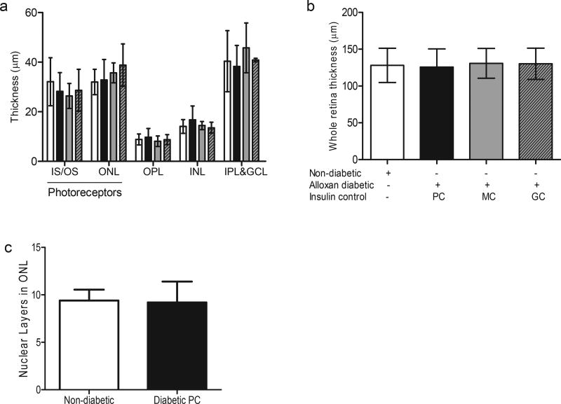
Fig. 1. Diabetes of 5 years duration does not result in photoreceptor cell loss in dogs.
Thickness of retinal regions (a), and the total retina (b) after 5 years of diabetes in dogs. Nuclear layers in the photoreceptor ONL were also compared between the non-diabetic and diabetic groups (c). All measurements were taken in the mid-retinal region of the nontapetal (inferior) nasal retina. There were no statistically significant differences noted between the diabetic and the non-diabetic group in all the data presented. n = 6 animals in the non-diabetic and diabetic PC groups, n = 3 for other groups. IS/OS, inner segment/outer segment of photoreceptors; ONL, outer nuclear layer (photoreceptors); OPL, outer plexiform layer; INL, inner nuclear layer; IPL&GCL, inner plexiform and ganglion cell layers. White bars are non-diabetic animals, black bars are alloxan diabetic animals with poor glycemic control, grey bars are alloxan diabetic animals with moderate insulin control, and grey bars with hatch lines are alloxan diabetic animals with good insulin control.

