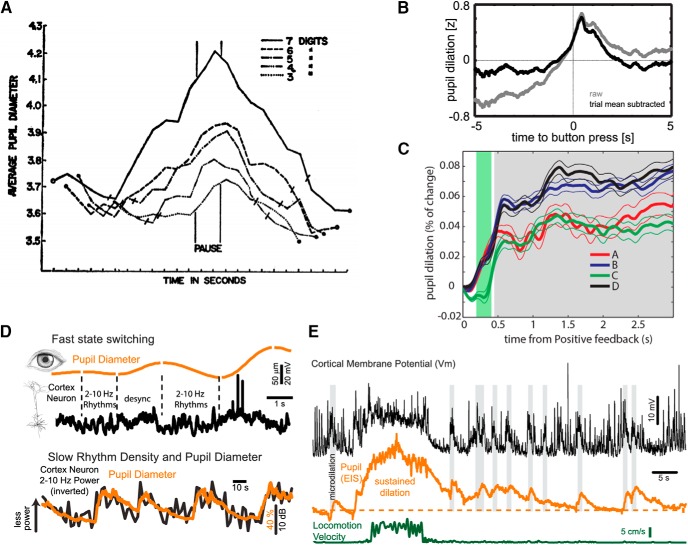
Figure 1.
Pupil diameter in human cognitive neuroscience and as an accurate predictor of rapid variations in parameters related to brain state and arousal in the mouse model. A, In a short-memory task, pupil dilation was modulated by the amount of items under active processing at any time: average pupil diameter (mm) for five subjects during auditory presentation (before “pause” period) and recall (after pause period) of digit strings of varying lengths (three to seven digits). The authors found that the pupil dilates when items are presented and constricts during report. The rate of change of these functions was related to task difficulty. From Kahneman and Beatty (1966). B. Pupil dilation departs from baseline ∼1 s before a volitional button press, peaks at 420 ms after the response, and relaxes back to baseline after ∼2 s, thus revealing the time of decision making. Authors interpreted pupil dilation as a marker of NE release from LC and as evidence for the latter role in consolidation of cognitive decisions. From Einhäuser et al. (2010). C, Pupil dilation displays an anticipatory response to uncertainty levels associated with options in a strategic gambling task (Iowa Gambling Task, IGT) where subjects are asked to maximize their profits by choosing the best drawing strategy from a set of four card decks, each of them delivering gains and losses following a pattern unknown for participants. Greater pupil dilation was observed in conditions with a low probability of incoming negative feedback (NF), as compared to conditions where NF had an enhanced probability to occur. Authors interpreted these results as evidence of pupil dilation signaling LC response to decision making in unfamiliar contexts. From Lavín et al. (2014). All axes in A–C depict pupil dilation (y-axis) versus time (x-axis). D, upper panel, Simultaneous recording of pupil diameter and membrane potential of a cortical neuron in layer 2/3 of the mouse primary visual cortex. Pupil diameter exhibits spontaneous variations in size even in the synchronized state and in the absence of locomotion. Note the strong relationship between slow (2–10 Hz) rhythmic synaptic activity and constriction, and the suppression of this activity with dilation. Lower panel, Comparison of pupil diameter and density of low-frequency (<10 Hz) rhythmic synaptic activity in a layer 5 pyramidal cell in the auditory cortex. Increases in pupil diameter are associated with prominent suppression of low-frequency synaptic activity (desynchronized state). From McGinley et al. (2015b). E, Whole-cell recordings from a layer 5 pyramidal neuron in auditory cortex of an awake mouse while simultaneously monitoring pupil diameter and locomotion. Brief dilations of the pupil (microdilations), are highlighted in gray and are associated with a suppression of the low-frequency activity and a depolarization of this neuron independent of locomotion. From McGinley et al. (2015b).

