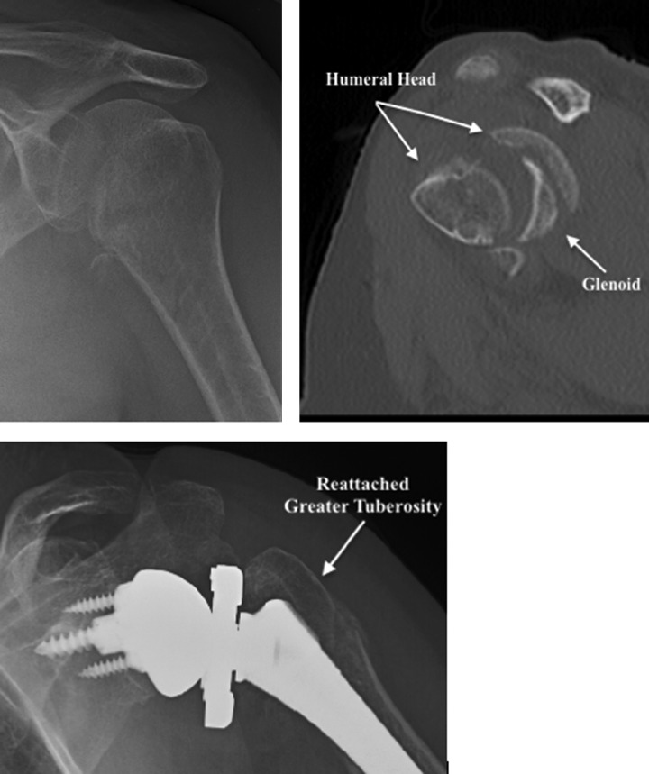
Figure 9.
Preoperative and postoperative views of a patient sustaining a comminuted head splitting fracture. Top left: AP view of the shoulder illustrating a fracture through the anatomic neck. Top right: sagittal CT slice clearly demonstrating the humeral head in multiple pieces. Bottom: Postoperative images of a reverse shoulder prosthesis illustrating the reattached greater tuberosity. AP denotes anteroposterior; CT, computed tomography.

