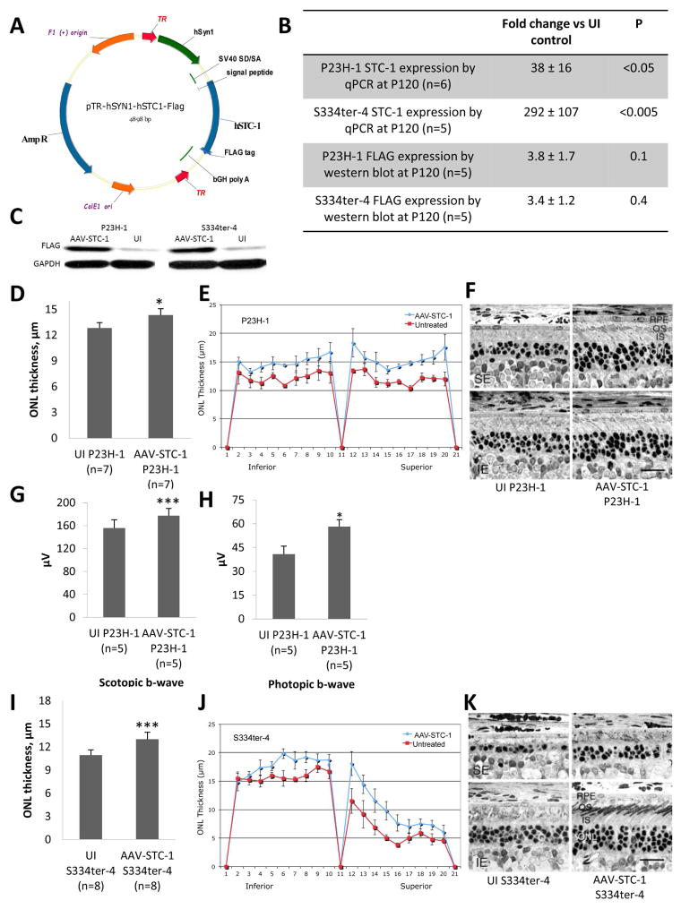
Fig. 1.
AAV-STC-1 construct and anatomic and functional rescue with AAV-STC-1. (A) AAV-STC-1 construct. Construct was designed to 1) incorporate a FLAG tag at the C-terminus, 2) maintain the signal peptide and 3) express hSTC-1 under the control of a retinal ganglion cell targeting promoter, human synapsin 1 (hSYN1). Vector was packaged in capsid modified AAV2 vector containing 3 surface-exposed tyrosine to phenylalanine mutations (AAV2-tripleYF). (B) After injection of AAV-STC-1 at P10, STC-1 gene expression was induced 38-fold in the P23H-1 rat (n=6, mean ± SEM, *P<0.05) and 292-fold in the S334ter-4 rat (n=5, mean ± SEM, ***P<0.005) at P120 while FLAG protein expression was induced 3.8-fold in the P23H-1 rat (n=5, P=0.1, Fig. 1B), and 3.4-fold in the S334ter-4 rat (n=5, P=0.4, Fig. 1B). (C) A representative western blot showing FLAG expression in AAV-STC-1 and UI retinas from P23H-1 and S334ter-4 rats. (D) At P120, AAV-STC-1 significantly improved ONL thickness of treated eyes of P23H-1 rats (n=7, mean ± SEM, *P<0.05). Note that this level of statistical significance was reached despite the fact that 3 of the 7 rats showed little rescue, presumably due to unsuccessful injections that occasionally occur with such experiments (LaVail et al., 1998) and variation of STC-1 overexpression between animals. (E) Spidergram reveals increased ONL thickness in superior and inferior retina of treated P23H-1 rats (n=3 showing greatest rescue; mean ± SEM; *P<0.05). (F) Histologic analysis revealed a thicker ONL and healthier photoreceptor inner (IS) and outer segments (OS) in eyes treated with AAV-STC-1 compared to those of the contralateral uninjected (UI) control eyes. The UI eyes are shown in the left column, and the injected eyes are shown on the right; the top row is taken from the superior equatorial (SE) retina, and the bottom row is taken from the inferior equatorial (IE) retina. Magnification bar = 25 μm. (G) At P120, scotopic (n=5, mean ± SEM, **P<0.005) and (H) photopic (n=5, mean ± SEM, *P<0.05) b-wave ERG response amplitudes were significantly increased in AAV-STC-1 treated P23H-1 animals. (I) At P120, AAV-STC-1 significantly improved ONL thickness of S334ter-4 rats (n=8, mean ± SEM, ***P<0.005). Note that this level of statistical significance was reached despite the fact that 2 of the 8 rats showed little or no rescue, presumably due to unsuccessful injections. (J) Spidergram reveals increased ONL thickness in superior and inferior retina in treated eyes of S334ter-4 rats (n=5 showing the greatest rescue; mean ± SEM; **P<0.01). (K) Histologic analysis shows a thicker ONL and healthier photoreceptor inner and outer segments in S334ter-4 eyes treated with AAV-STC-1 compared to those of the contralateral uninjected (UI) control eye. Light micrographs represent the most extensive rescue in this cohort. The labels are the same as in (F).

