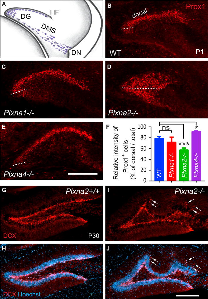
Figure 1. Plxna2 and Plxna4 Regulate GC Distribution in the Developing DG.
(A) In the developing mouse DG, immature GCs (blue cells) migrate from the dentate notch (DN) of the telencephalic neuroepithelium, along the dentate migratory stream (DMS) toward the hippocampal fissure (HF).
(B–E) Anti-Prox1 staining of coronal brain sections through the rostral pole of the P1 hippocampus of (B) WT (n = 4), (C) Plxna1−/− (n = 3), (D) Plxna2−/− (n = 5), and (E) Plxna4−/− (n = 3) pups. The border between the dorsal and ventral GCL is marked by a dotted line through the medial pole of the DG.
(F) Quantification of Prox1+ cells in the dorsal blade of the GCL compared to total Prox1 (GCL + hilus) labeling. Results are shown as mean value ± SD. *p < 0.05; ***p < 0.001, by one-way ANOVA, with Dunn post hoc t test. ns, not significant.
(G–J) Coronal sections through the dorsal hippocampus of P30 (G and H) Plxna2+/+ (n = 3) and littermate (I and J) Plxna2−/− mice (n = 3) stained with anti-DCX (G and I) and with anti-DCX + Hoechst dye 33342 (H and J). Arrows point to ectopically positioned DCX+ cells in the dentate molecular layer. Scale bar, 200 µm.

