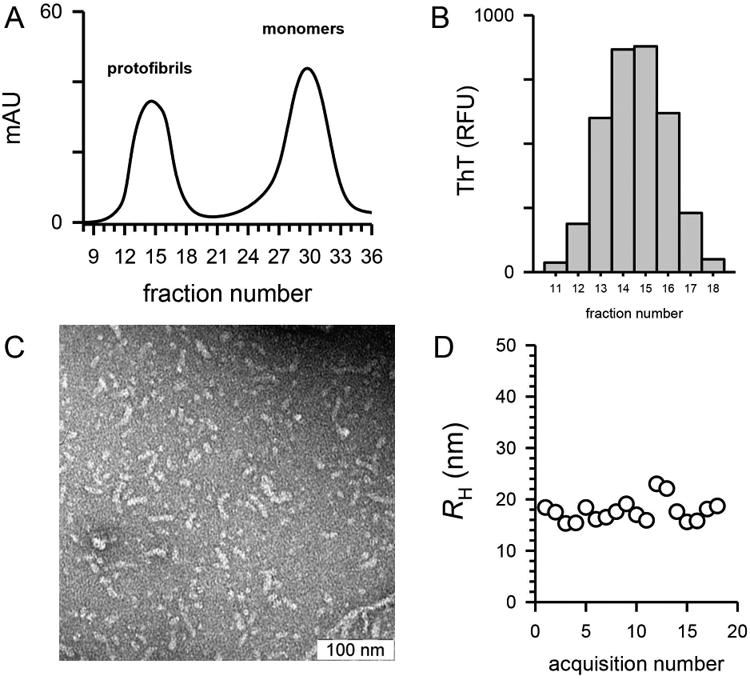
Figure 1.

Characterization of Aβ42 protofibrils used for immunization. Panel A. Aβ42 protofibrils were generated as described in the Methods and isolated on a Superdex 75 SEC column in aCSF. UV absorbance at 280 nm was monitored during the elution (solid line). Panel B. Protofibril SEC fractions (0.5 ml) were diluted by 10 into aCSF containing 5 μmol/L ThT and fluorescence emission was measured as described in the Methods. Panel C. Fractions 13-16 were pooled to yield 2 mL of 42 μmol/L protofibrils. A sample (10 μl) was applied to a copper formvar grid, and imaged by TEM at a magnification of 50,000. The scale bar represents 100 nm. Panel D. Mean RH was measured by DLS of the Aβ42 protofibril pool and a plot of RH vs acquisition number is shown.
