
Figure 2.
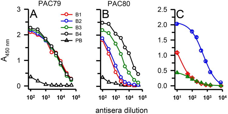
Serum antibody titer determination for Aβ42 protofibrils. Serum from a pre-bleed (PB) and multiple bleeds (B1-4) of two rabbits (PAC79, Panel A; PAC80, Panel B) immunized with Aβ42 protofibrils was analyzed via indirect ELISA. 96-well ELISA plates were coated with Aβ42 protofibrils (18 ng/well) and treated with increasing dilutions of anti-serum and anti-rabbit IgG-HRP. Each data point represents the average of n=4 trials. Panel C. Varying dilutions of serum antibody were used to detect 18 ng/well of Aβ42 protofibrils (blue circles), Aβ42 monomers (red diamonds), or Aβ42 fibrils (green triangles) by indirect ELISA. Data points (± standard error measurement, SEM) represents the average of n=3 trials and were fit to a 3-parameter hyperbolic decay equation using SigmaPlot software.