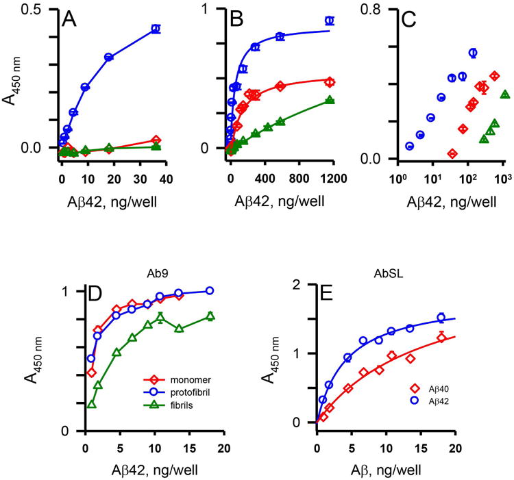
Figure 3.

AbSL antiserum displays selectivity for Aβ42 protofibrils. 96-well ELISA plates were coated with varying amounts of Aβ42 protofibrils, Aβ42 monomers, and Aβ42 fibrils (0.5-1156 ng/well) and analyzed by indirect ELISA with AbSL anti-serum (1:10,000 dilution) and anti-rabbit IgG-HRP (Panels A-C). Panel A depicts the lower Aβ42 concentrations and Panel B shows the extended Aβ42 concentration range. Data in Panel B were fit to a 3-parameter single rectangular hyperbola equation using SigmaPlot software. Data points (± SEM) represent the average of n=3 trials. Panel C is a semi-log re-plot of the central data from Panel B. Panel D. Plates coated with Aβ42 protofibrils, Aβ42 monomers, and Aβ42 fibrils were treated with the N-terminal Aβ antibody Ab9 (1:5,000 dilution) and anti-mouse IgG-HRP. Panel E. Aβ42 protofibrils and Aβ40 protofibrils were prepared separately by different protocols and isolated by SEC. 96-well ELISA plates were coated with a concentration range of protofibrils (0.9-18 ng per well) and analyzed by indirect ELISA. Wells were incubated with AbSL (1:1,000 dilution) followed by anti-rabbit IgG-HRP secondary antibody. Curve-fitting of the data points was performed using SigmaPlot software. Error bars depict SEM for n = 3 wells.
