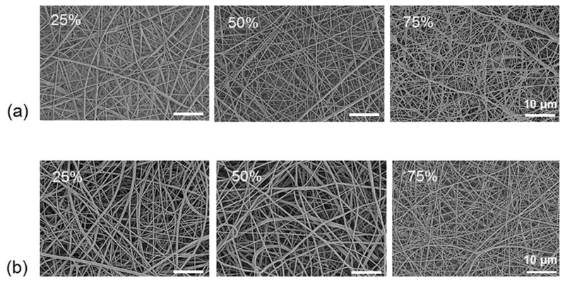
Fig. 2.
Scanning electron microscopy (SEM) images of fibrous membranes. AT-PCL was blended at different w/w ratios (25%, 50%, 75%) with high molecular weight PCL and processed by electrospinning. For all AT-PCL to PCL ratios, free standing membranes were produced. (a) pristine AT-PCL membranes, (b) phytic acid doped AT-PCL membranes.

