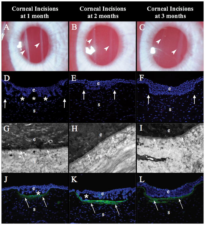
Figure 3.
Rabbit corneas at one, two and three months after non-perforating linear incisions. (A) At one month after the surgery, the two incisions (arrowheads) were visible as dense linear opacities in all nine corneas that reached this time point (mag 25X). (B) At two months after surgery, the incisions (arrowheads) were less opaque in all six corneas that reached this time point (mag 25X). (C) At three months after surgery, the incisions (arrowheads) were faint compared to the two-month time point in all three corneas that reached this time point (mag 25X). (D), (E), and (F) At one, two or three months after surgery, respectively, there were no alpha-smooth muscle actin (α-SMA)+ cells anywhere in the cornea, including around the epithelial plugs (arrows), in three corneas at each time point (mag 200X). (G), (H), and (I) At one, two and three months, respectively, after surgery the EBM (arrows), including lamina lucida and lamina densa, were fully regenerated and surrounded the epithelial plugs in all three corneas at each time point (mag 23,000X). (J) At 1 month after surgery, both incision in each of the three corneas studied had collagen type III (COL3, arrows) deposited in the subepithelial stroma beneath the epithelial plugs (mag 200X). At two months (K) and three months (L) after surgery, more COL3 deposition (arrows), compared to the one month time point, was noted in the subepithelial stroma surrounding the epithelial plugs in each of the three corneas at each time points. Blue is DAPI staining of cell nuclei; (e) epithelium; (s) stroma; (*) indicates artifact separation of the epithelium from the stroma that occurs during cutting of the section.

