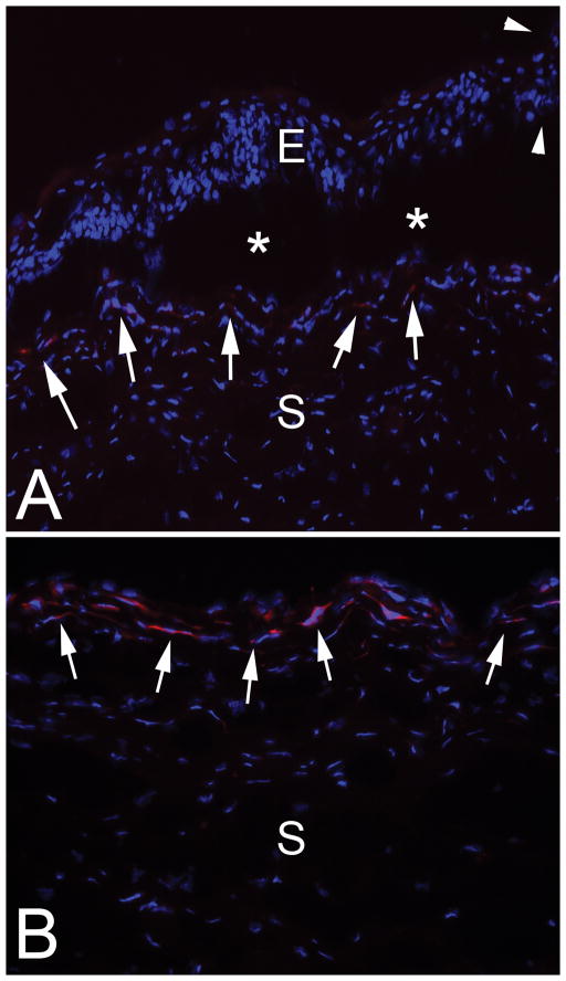
Fig. 2.
Immunohistochemistry for the alpha-smooth muscle actin (α-SMA) marker for myofibroblasts in the cornea of rabbit #1. Blue stain is DAPI that stains all cell nuclei. A. The epithelial leading edge (arrowheads) is rolled and the epithelium for up to 0.3 mm posterior to the leading edge showed poor adhesion and repeated artifactual dissociation (*) from the underlying stroma in all sections cut with a cryostat. α-SMA+ myofibroblasts are present at the stromal surface all along the dissociated epithelium (arrows) and, therefore, peripheral to the leading-edge of the epithelium in the PED. E is epithelium and S is stroma. Magnification 400X. B. In the center of the PED the stromal surface has prominent α-SMA+ myofibroblasts (arrows). S is stroma. Mag. 400X

