
Figure 2.
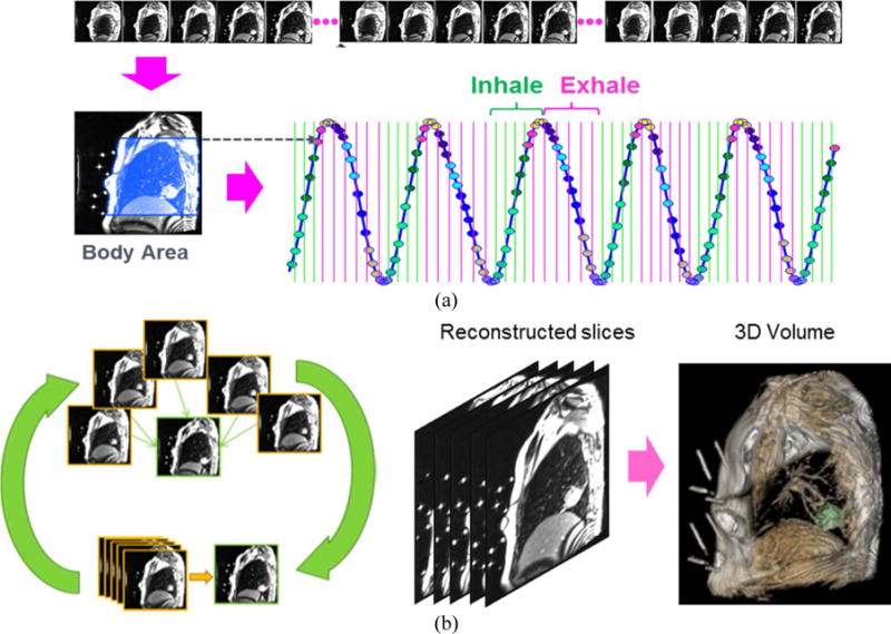
The 4D-MRI reconstruction process. (a) Phase binning based on body area. The blue region on the left image shows the automatically computed body area including the lung. (b) 4D-MRI reconstruction by groupwise registration. For each breathing phase, each slice image is reconstructed by groupwise registration of multiple sorted images (left). Slice reconstruction is repeated for all slices, and a 3D reconstruction at each breathing phase is computed by stacking the reconstructed slice images (middle). Tumor is then segmented from the reconstructed 4D-MRI to form a tumor template (right, green indicates the segmented tumor).