
Figure 4.
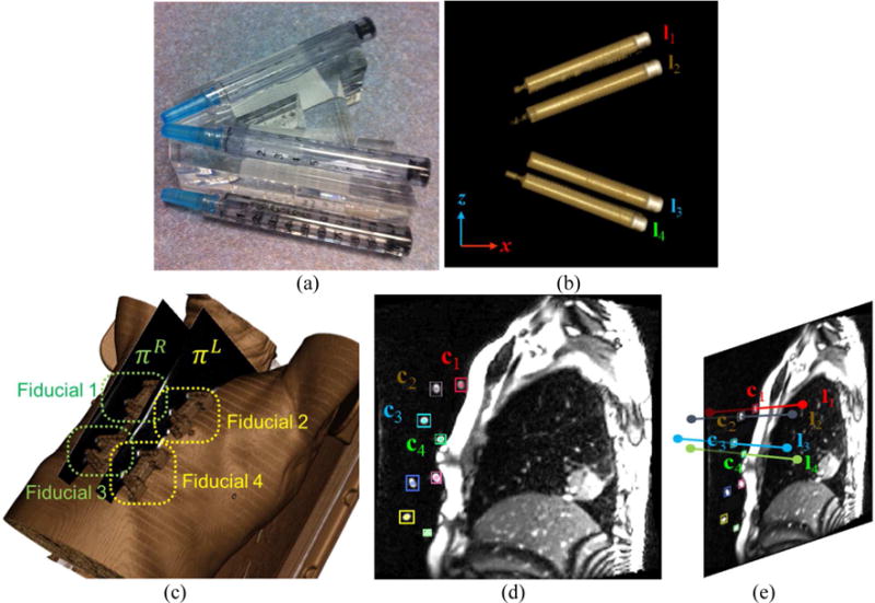
Fiducial tracking. (a) The photograph of the fiducial, composed of 4 cylinders filled with normal saline. (b) Volume rendering of the fiducial (cylinders only). (c) Locations of four fiducials on volumetric rendering. (d) Fiducial segmentation in 2D MR image (colored boxes). (e) 3D fiducial tracking by matching 3D fiducial model (4 line segments) to the segmented fiducial markers shown in (d).