
Figure 2.
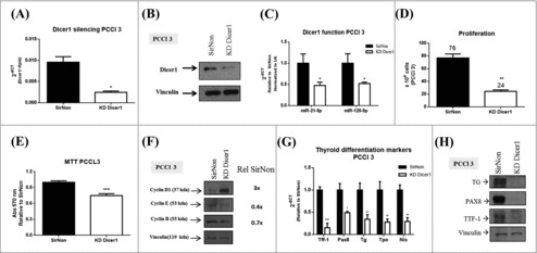
Silencing of Dicer1 in PCCl 3 cells. Cells were transfected with nonsilencing control siRNA (SirNon) or specific Dicer1 siRNA (KD Dicer1) and (A) Dicer1 mRNA and (B) protein levels were assessed by q-RT-PCR and western blot, respectively. (C) Expression of miR-21-5p and miR-125-5p to evaluate Dicer1 function. (D) Number of PCCl 3 cells was counted using trypan blue 48 hours post transfection with SirNon or specific Dicer1 siRNA. (E) In parallel, cell viability was assessed using MTT assay in the same conditions. (F) Expression of cyclin D1, E and B by western blot 48 hours after the transfection of PCCl 3 cells with SirNon or specific Dicer1 siRNA. Densitometry intensity quantification was calculated as ratio target: vinculin and relative to SirNon. (G) mRNA levels of the thyroid differentiation markers Ttf-1, Pax8, Tg, Tpo and Nis 48 hours after the transfection of PCCl 3 cells with SirNon or specific Dicer1 siRNA were evaluated by q-RT-PCR. (H) Protein levels of thyroglobulin (TG), PAX8 and TTF-1 were assessed by western blot in the same conditions. Data were relative to SirNon. Vinculin was used as loading control for western blot. Rpl4 and pseudogene U6 were used as housekeeping for q-RT-PCR of mRNA and miR levels, respectively. p < 0.05, p < 0.01; p < 0.001.