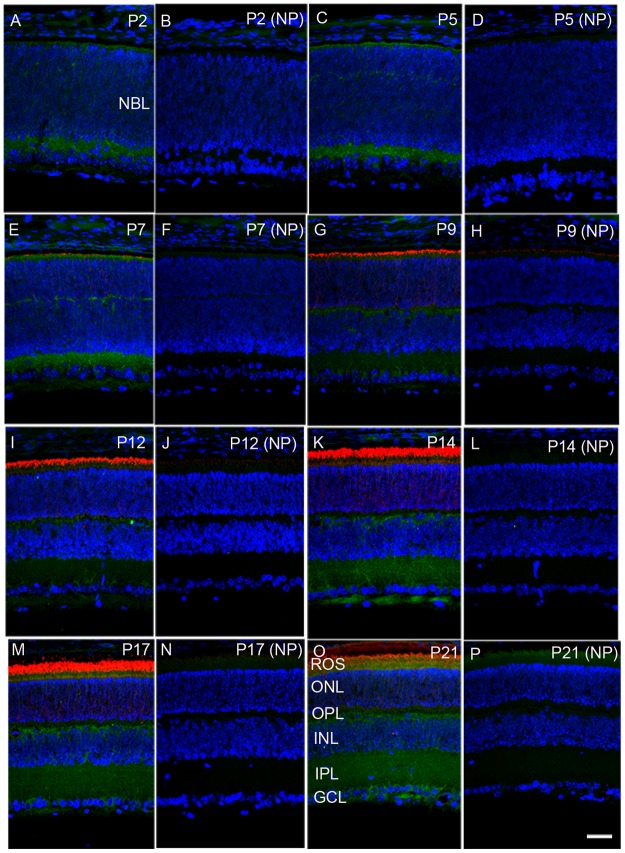
Figure 1. Developmental expression of PP2A in the retina.
Mouse retinal sections were prepared on postnatal day (P) 2, P5, P7, P9, P12, P14, P17, and P21 and were stained with PP2A (green) and rhodopsin (red). Panels A, C, E, G, I, K, M, and O are stained with primary antibodies, whereas panels B, D, F, H, J, L, N, and P are no-primary controls. NBL, neuoblastic layer; ROS, rod outer segments; ONL, outer nuclear layer; OPL, outer plexiform layer; INL, inner nuclear layer; IPL, inner plexiform layer; GCL, ganglion cell layer. Scale bar = 50 μm.

