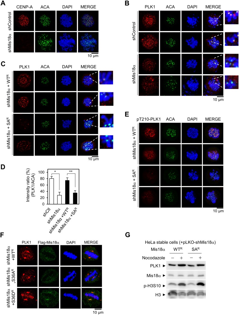
Figure 4. Mis18α phosphorylation enhances PLK1 kinetochore recruitment.
(A) HeLa cells were infected with lentivirus expressing either control shRNA (shControl) or shRNA against Mis18α (shMis18α). Cells were fixed at prometaphase by releasing for 30 min after monastrol treatment and stained with anti-ACA (centromere marker) or anti-CENP-A antibody. Confocal image with 1,000× magnification. (B) Cells prepared as in A were co-stained with anti-PLK1 and anti-ACA antibodies. (C) HeLa cells stably expressing shRNA-resistant form of Mis18α (WTR and SAR) were infected with lentivirus expressing shMis18α. Cells were co-stained with anti-PLK1 and anti-ACA antibodies at prometaphase. (D) The number of cells showing high intensity of PLK1 staining, ACA signal as a control (PLK1/ACA), from B and C was presented in percentage. P value is calculated by t-test (*p < 0.05, **p < 0.01). (E) pThr210-PLK1 was co-stained with ACA in the same cells as C. (F) HeLa cells stably expressing shRNA-resistant form of Mis18α (WTR, SAR and SDR) were co-stained with anti-PLK1 and anti-Flag Mis18α antibody at metaphase. (G) Immunoblot for PLK1 level in reconstituted HeLa stable cell lines.

