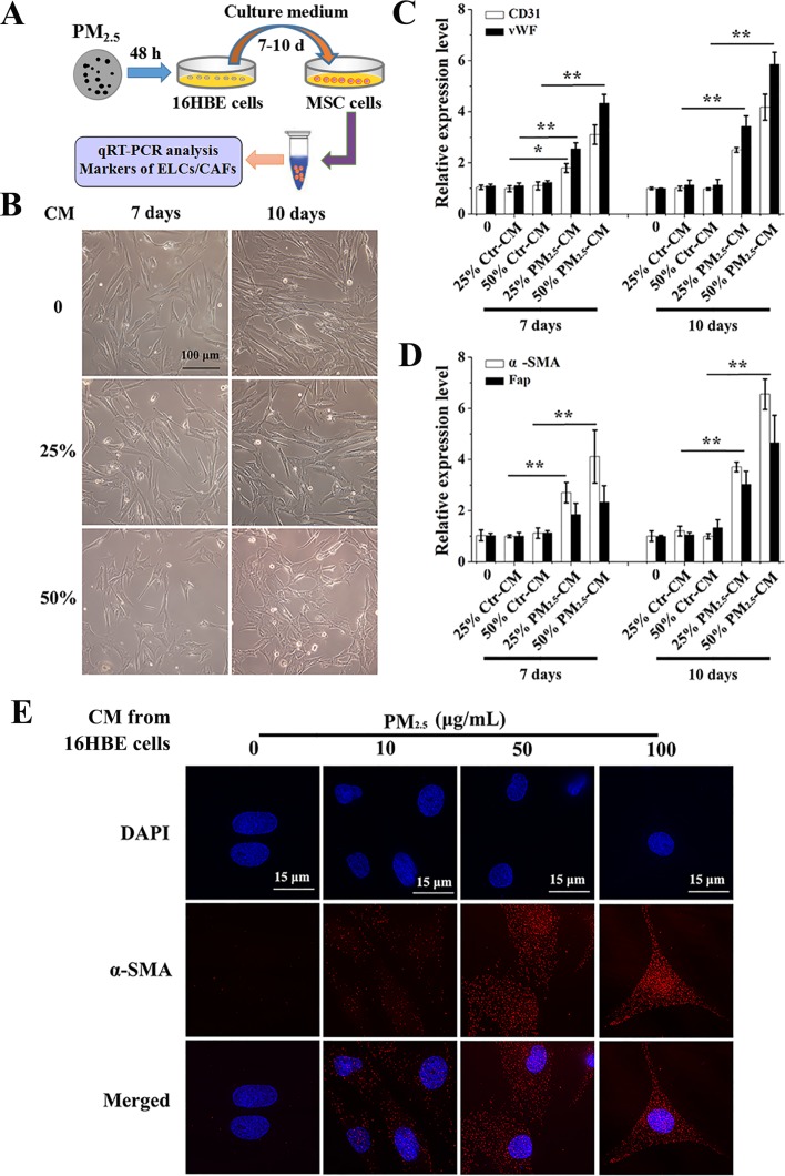
Figure 2. Conditioned medium from PM2.5-treated 16HBE cells induces the differentiation of BMSCs.
(A) Experimental protocol for PM2.5-exposed cells. 16HBE cells were treated with PM2.5 (10–100 μg/mL) for 48 hours, cell culture supernatants were removed, centrifuged and diluted 1:4 or 1:2 with DMEM/HBMSC-GM medium (without serum). BMSCs were exposed to CM from 16HBE cells for 7 or 10 days. (B) The morphology of BMSCs. mRNA levels of (C) CD31, vWF, (D) a-SMA and Fap were measured. (E) The localization of a-SMA in BMSCs upon 50% CM exposure for 7 days were determined (60× magnification); scale bars = 15 μm. The values were showed as means ± SD of triplicate determinations. *p < 0.05, **p < 0.01, compared with control.

