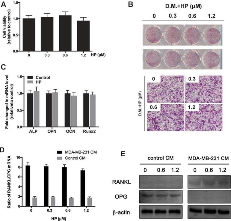
Figure 3. Hypericin has minimal effects on breast-cancer-cell induced increase in RANKL/OPG ratio in osteoblast.
(A) MC3T3-E1 cells were treated with the indicated concentrations of HP for 48 h. Cell viability was measured by the CCK-8 assay and is presented as percentages relative to the control group. (B) MC3T3-E1 cells were incubated in an osteogenic differentiation medium (DM) containing the indicated concentrations of HP. On day 14, differentiated cells were stained with ALP to detect the osteoblastic differentiation ability. Data represent the results of one of three experiments with similar results. (C) MC3T3-E1 cells were incubated with 1.2 μM HP for 7 days. The mRNA expression of ALP, OPN, OCN, and Runx2 genes was determined by real-time PCR and presented as ratios relative to the control group. All expression levels were normalized to GAPDH mRNA levels in corresponding samples. Data represent the means ± SD of three independent experiments (*P < 0.05). (D) Effects of HP on breast cancer cell-induced increase in RANKL/OPG ratio in osteoblasts. MDA-MB-231 cells (2 × 10⁶ cells/well) were treated with the indicated doses of HP for 24 h. A conditioned medium (CM) was harvested. In addition, bone marrow stromal cells (BMSCs; 1 × 10⁵ cells/well) were seeded onto 24-well plates and induced to differentiate via addition of ascorbic acid (50 g/mL), β-glycerophosphate (10 mM), and dexamethasone (10−8 M) for 7 days. Then, they were stimulated with CM for another 24 h. Total RNA was collected and subjected to quantitative real-time PCR using the indicated primers. (E) Effects of HP on breast cancer cell-induced RANKL and OPG protein expression level. MDA-MB-231 cells (2 × 10⁶ cells/well) were treated with the indicated doses of hypericin for 24 h. CM was harvested. Separately, BMSCs (1 × 10⁵ cells/well) were seeded onto 6-well plates and induced to differentiate via addition of ascorbic acid (50 g/mL), β-glycerophosphate (10 μM), and dexamethasone (10−8 M) for 7 days. Then, they were stimulated with CM for another 24 h. Whole cell extracts were prepared and subjected to western blot analysis as described in the materials and methods.

