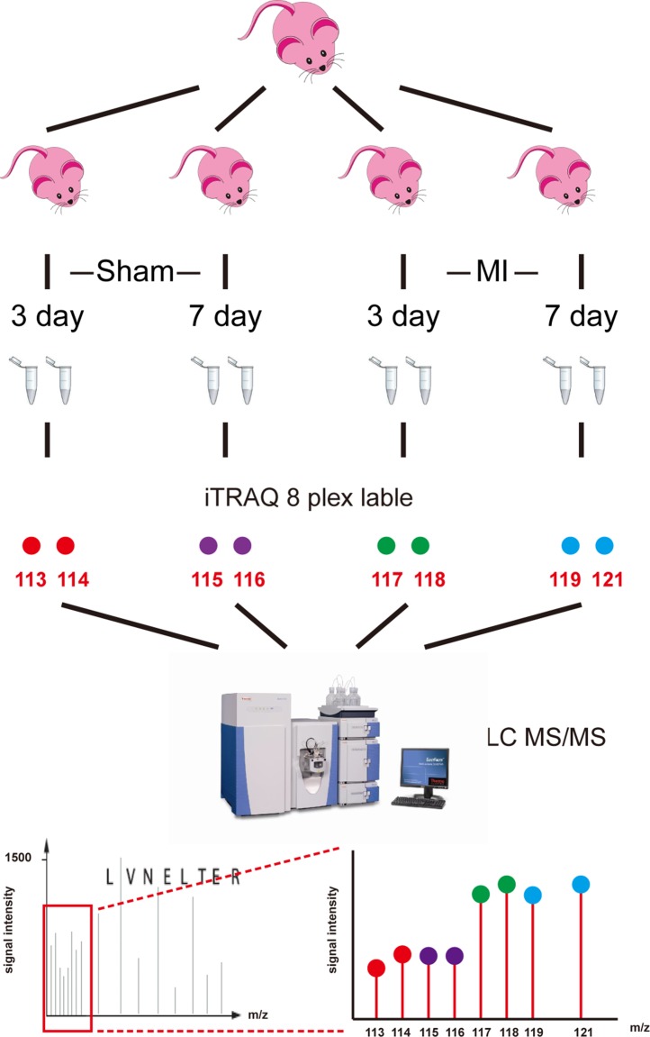
Figure 1. Workflow of iTRAQ analysis of acute myocardial infarction mouse model.
Left ventricles were obtained from acute myocardial infarction model mice at 3 days and 7 days after surgery (MI-3d and MI-7d) and sham operation (Sham-3d and Sham-7d). Eight independent biological duplicates were used for each group. Pooled protein samples were digested by trypsin. Two technical replicates were performed for each group (isobaric tags 113 and 114 for sham-3d; 115 and 116 for sham-7d; 117 and 118 for MI-3d; 119 and 121 for MI-7d). iTRAQ-labeled peptides were subsequently fractionated by HPLC and then were analyzed by LC-MS/MS.

