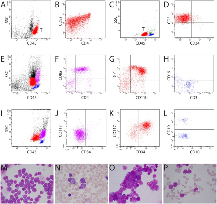
Figure 2. Different locations of blasts from murine and human T-ALL.
(A) Murine lymphoblasts from T-ALL (red population) located in a distinctive “blast gate” (aBG, CD45bright). T = T-lymphoblasts. (B) Murine lymphoblasts expressing CD4, CD8, and CD3 (data not shown). A, B: mouse #1364. (C) Human T-ALL blasts (red population) were present in the cBG (CD45dim). Blue population: lymphocytes; green population: monocytes. (D) Expression of cyCD3 on human blasts. C, D: patient LB. (E) Myeloblasts (red population) and T-ALL blasts (purple population) were located in different regions in the bone marrow of mouse #1329 (E-H). (F) T-ALL blasts expressing CD4/CD8. (G) Expression of CD11b and Gr1 on myeloblasts. (H) Mature lymphocytes expressing either CD3 or CD19. (I-L) Location of monoblasts in the aBG (purple population) from patient TK with AML M4. Note the slightly higher CD45 fluorescence intensity of monoblasts than of the mature lymphocytes (I). Monoblasts were negative for CD117 or CD34 (J), whereas myeloblasts (red population) expressed both CD117 and CD34 (K). (L) Mature lymphocytes expressed CD19 (not CD10) or CD3 (data not shown). (M, N) Bone marrow cytospin and blood smear showing predominant murine and human lymphoblasts in mouse #1364 (A, B) and patient LB (C, D), respectively. (O) Bone marrow cytospin showing infiltration of myeloblasts and lymphoblasts in the bone marrow of mouse #1329 (E-H). (P) Blood smear showing blasts and promonocytes in patient TK with AML M4 (I-L). Blasts were positive for Sudan Black B and non-specific esterase (Supplementary Figure 3).

