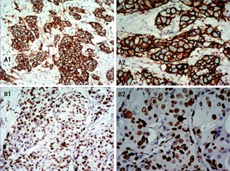
Figure 1. Positive expression of HER-2 and Ki-67 in invasive breast cancer as shown by IHC.
Positive HER-2 expression: the cell membrane is brown and continuous. (A1) 100x magnification, (A2) 200x magnification. High Ki-67 expression: the nucleus is brown, with tumor cell positivity in 70% of the cells, (B1) 100x magnification, (B2) 200x magnification. IHC: immunohistochemistry.

