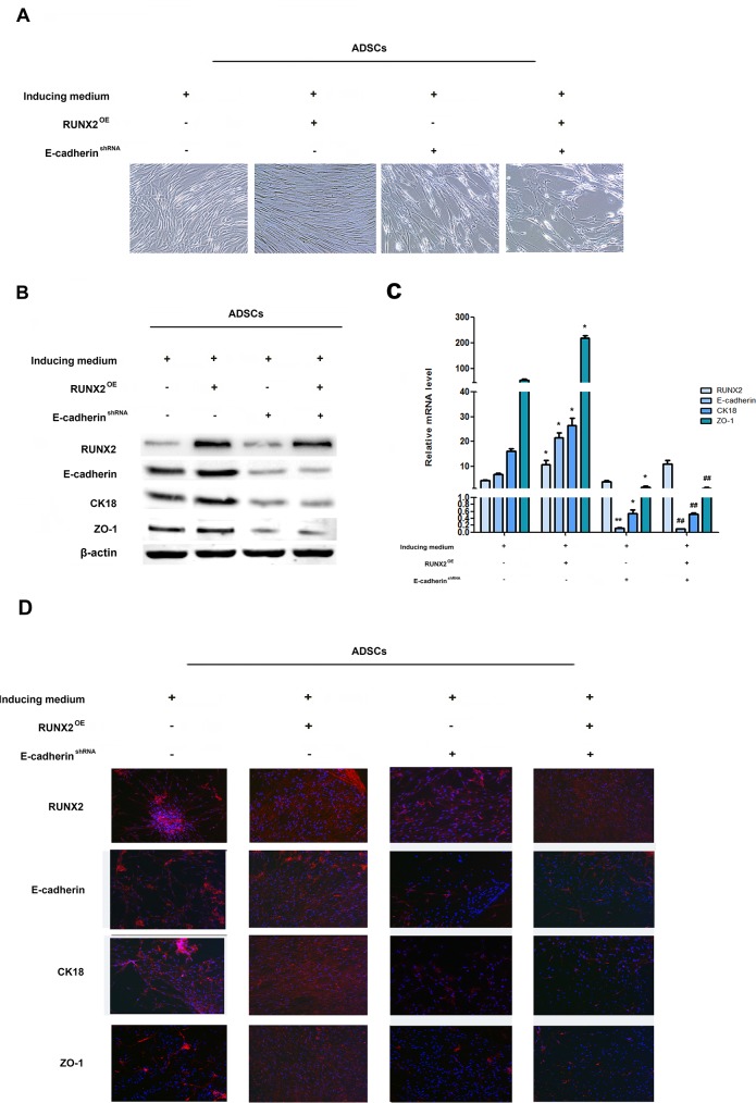
Figure 4. RUNX2 promotes ADSCs differentiation through E-cadherin.
(A) Representative image of ADSC morphology under the microscope after ADSCs given LV5-RUNX2 or LV3-shE-cadherin (×200). (B) Western blot analyses of protein levels of RUNX2, E-cadherin, CK18 and ZO-1 after ADSCs given LV5-RUNX2 or LV3-shE-cadherin. β-actin was used as an internal control. (C) qRT-PCR analyses of mRNA levels of E-cadherin, CK18 and ZO-1 after ADSCs given LV5-RUNX2 or LV3-shE-cadherin. (D) Immunofluorescence analysis to assess the expression of RUNX2, E-cadherin, CK18 and ZO-1 after ADSCs given LV5-E-cadherin or LV3-shE-cadherin (×100). Nuclei are stained by 4,6-diamidino-2-phenylindole. These data are expressed as the mean ± SD (*P<0.05, **P<0.01versus induced medium group; #P<0.05, ##P<0.01 versus LV5-RUNX2 group). Each experiment was repeated at least three times.

