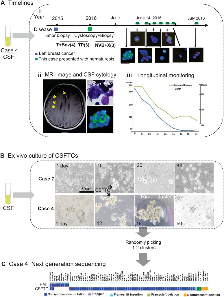
Figure 1. Overview of SE-i•FISH platform for dynamic monitoring and ex vivo culture of CSFTCs.
(A) The treatment timelines received by the patient (NM04). (i) From 2015 until 2016, the patient received four cycles of adjuvant T+Bev (Paclitaxel/Bevacizumab) and three cycles of TP (Paclitaxel/DDP) chemotherapy. In January 2016, the patient presented with hematuresis. Following urethrocystoscopy biopsy, the patient received three cycles of NVB+X (Vinorelbine/Capecitabine). The treatment had to stop as she presented with excruciating headache and vomiting. On the basis of the diagnosis of leptomeningeal metastases by MRI and CSF cytology, the patient received regularly intrathecal methotrexate and cytarabine. Green arrows mean intrathecal chemotherapies. The immunofluorescent staining for cytokeratin (CK, green), chromosome 8 (Yellow), CD45 (Red), and nuclei (DAPI, blue). (ii) Representative images of MRI and CSF cytology. (Upper right panel) Light microscopic imaging with Papanicolaou staining. (iii) The number of CSFTCs and the intracranial pressure are shown in the right panel. (B) Ex vivo culture of CSFTCs. 5–10 ml CSF were obtained from patients and then enriched. CSFTCs were cultured in a medium without serum under suspension culture conditions. The CSFTCs of NM04 and NM07 could be expanded in short term as indicated. Scale bar, 50 μm. (C) At Day 18, the tumor clusters of case NM04 were randomly picked. The exome sequencing was performed on DNA extracted from CSFTCs and the paired primary tumor. The distribution of somatic mutations detected by exome sequencing is presented in a heat map.

