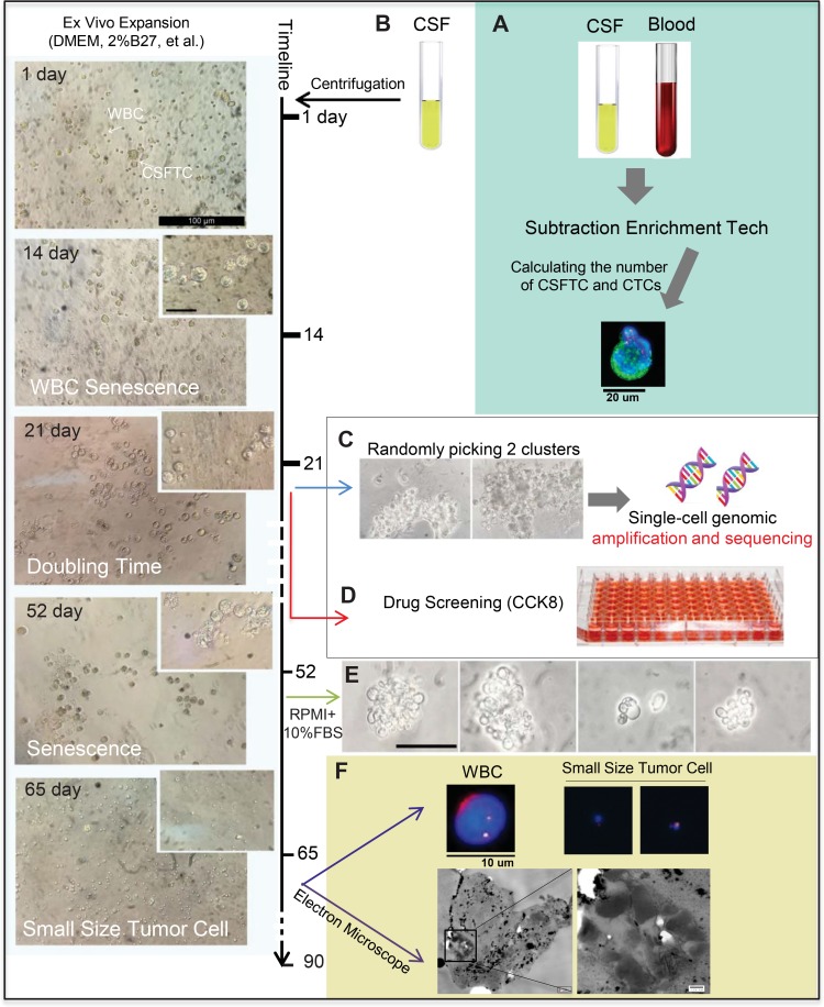
Figure 3. Ex vivo expansion of CSFTCs from case NM01.
The left panel showed representative images of ex vivo culture of CSFTCs. The CSFTCs could be expanded for more than seven weeks, but after 90 days they gradually underwent senescence. (A) The number of CTCs in blood and CSFTCs were recorded by the SE-i•FISH platform. (B) Cerebrospinal fluid was collected and centrifuged. Subsequently, CSFTCs were enriched by subtraction enrichment (SE) technology. (C) At Day 21, cultures appeared tumor clusters, and then two tumor clusters were picked for single-cell genomic amplification and exome sequencing. (D) Ex vivo drug sensitivity test was assessed by CCK8 assay. (E) The morphological changes of CSFTCs were observed, when the special non-serum medium was switched to the medium with 10% FBS. At Day 65, we observed the small size cells. Representative images of immunofluorescence (up) and scanning electron microscope (down) are shown in (F).

