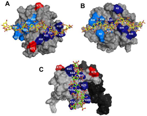
Fig. 2.

Models of heparin bound CXCL1 and CXCL5 complexes. Panels A and B shows the non-overlapping heparin-binding surfaces in CXCL1 (defined as α- and β-domains). Heparin-binding residues from both monomers are highlighted in light and dark blue and residue K71 from both monomers are labelled in red. Panel C show the heparin-binding surface in CXCL5. Heparin binding residues were highlighted in blue and residue K76 is shown in red. The second monomer of the dimer is shown in black for clarity.
