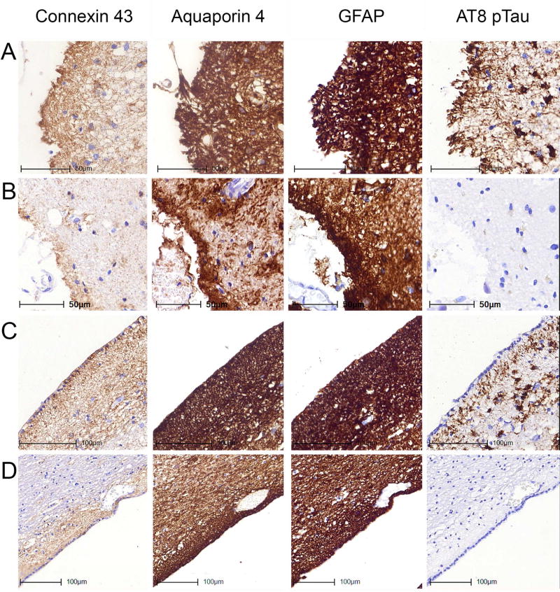
Fig 5.
Immunostaining patterns of Cx43, AQP4, GFAP and AT8 pTau in the subpial location of the inferior temporal gyrus (A, B) and the subependymal location of the inferior horn of the lateral ventricle (C, D) representing different degrees of tau pathologies. Images A and C represent case ARTAG-2 demonstrating subpial and subependymal ARTAG; B and D represent case CO-8 lacking subpial and subependymal ARTAG.

